Recommendation
Polyphenisms offer an opportunity to study the links between phenotype, development, and environment in a controlled genomic context (Simpson, Sword, & Lo, 2011). In organisms with short generation times, individuals living and developing in different seasons encounter different environmental conditions. Adaptive plasticity allows them to express different phenotypes in response to seasonal cues, such as temperature or photoperiod. Such phenotypes can be morphological variants, for instance displaying different wing patterns as seen in butterflies (Brakefield & Larsen, 1984; Nijhout, 1991; Windig, 1999), or physiological variants, characterized for instance by direct development vs winter diapause in temperate insects (Dalin & Nylin, 2012; Lindestad, Wheat, Nylin, & Gotthard, 2019; Shearer et al., 2016).
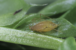
Many aphids display cyclical parthenogenesis, a remarkable seasonal polyphenism for reproductive mode (Tagu, Sabater-Muñoz, & Simon, 2005), also sometimes coupled with wing polyphenism (Braendle, Friebe, Caillaud, & Stern, 2005), which allows them to switch between parthenogenesis during spring and summer to sexual reproduction and the production of diapausing eggs before winter. In the pea aphid Acyrthosiphon pisum, photoperiod shortening results in the production, by parthenogenetic females, of embryos developing into the parthenogenetic mothers of sexual individuals. The link between parthenogenetic reproduction and sexual reproduction, therefore, occurs over two generations, changing from a parthenogenetic form producing parthenogenetic females (virginoparae), to a parthenogenetic form producing sexual offspring (sexuparae), and finally sexual forms producing overwintering eggs (Le Trionnaire et al., 2022).
The molecular basis for the transduction of the environmental signal into reproductive changes is still unknown, but the dopamine pathway is an interesting candidate. Form-specific expression of certain genes in the dopamine pathway occurs downstream of the perception of the seasonal cue, notably with a marked decrease in the heads of embryos reared under short-day conditions and destined to become sexuparae. Dopamine has multiple roles during development, with one mode of action in cuticle melanization and sclerotization, and a neurological role as a synaptic neurotransmitter. Both modes of action might be envisioned to contribute functionally to the perception and transduction of environmental signals.
In this study, Le Trionnaire and colleagues aim at clarifying this role in the pea aphid (Le Trionnaire et al., 2022). Using quantitative RT-PCR, RNA-seq, and in situ hybridization of RNA probes, they surveyed the timing and spatial patterns of expression of dopamine pathway genes during the development of different stages of embryo to larvae reared under long and short-day conditions, and destined to become virginoparae or sexuparae females, respectively. The genes involved in the synaptic release of dopamine generally did not show differences in expression between photoperiodic treatments. By contrast, pale and ddc, two genes acting upstream of dopamine production, generally tended to show a downregulation in sexuparare embryo, as well as genes involved in cuticle development and interacting with the dopamine pathway. The downregulation of dopamine pathway genes observed in the heads of sexuparare juveniles is already detectable at the embryonic stage, suggesting embryos might be sensing environmental cues leading them to differentiate into sexuparae females.
The way pale and ddc expression differences could influence environmental sensitivity is still unclear. The results suggest that a cuticle phenotype specifically in the heads of larvae could be explored, perhaps in the form of a reduction in cuticle sclerotization and melanization which might allow photoperiod shortening to be perceived and act on development. Although its causality might be either way, such a link would be exciting to investigate, yet the existence of cuticle differences between the two reproductive types is still a hypothesis to be tested. The lack of differences in the expression of synaptic release genes for dopamine might seem to indicate that the plastic response to photoperiod is not mediated via neurological roles. Yet, this does not rule out the role of decreasing levels of dopamine in mediating this response in the central nervous system of embryos, even if the genes regulating synaptic release are equally expressed.
To test for a direct role of ddc in regulating the reproductive fate of embryos, the authors have generated CrispR-Cas9 knockout mutants. Those mutants displayed egg cuticle melanization, but with lethal effects, precluding testing the effect of ddc at later stages in development. Gene manipulation becomes feasible in the pea aphid, opening up certain avenues for understanding the roles of other genes during development.
This study adds nicely to our understanding of the intricate changes in gene expression involved in polyphenism. But it also shows the complexity of deciphering the links between environmental perception and changes in physiology, which mobilise multiple interacting gene networks. In the era of manipulative genetics, this study also stresses the importance of understanding the traits and phenotypes affected by individual genes, which now seems essential to piece the puzzle together.
References
Braendle C, Friebe I, Caillaud MC, Stern DL (2005) Genetic variation for an aphid wing polyphenism is genetically linked to a naturally occurring wing polymorphism. Proceedings of the Royal Society B: Biological Sciences, 272, 657–664. https://doi.org/10.1098/rspb.2004.2995
Brakefield PM, Larsen TB (1984) The evolutionary significance of dry and wet season forms in some tropical butterflies. Biological Journal of the Linnean Society, 22, 1–12. https://doi.org/10.1111/j.1095-8312.1984.tb00795.x
Dalin P, Nylin S (2012) Host-plant quality adaptively affects the diapause threshold: evidence from leaf beetles in willow plantations. Ecological Entomology, 37, 490–499. https://doi.org/10.1111/j.1365-2311.2012.01387.x
Le Trionnaire G, Hudaverdian S, Richard G, Tanguy S, Gleonnec F, Prunier-Leterme N, Gauthier J-P, Tagu D (2022) Dopamine pathway characterization during the reproductive mode switch in the pea aphid. bioRxiv, 2020.03.10.984989, ver. 4 peer-reviewed and recommended by Peer Community in Zoology. https://doi.org/10.1101/2020.03.10.984989
Lindestad O, Wheat CW, Nylin S, Gotthard K (2019) Local adaptation of photoperiodic plasticity maintains life cycle variation within latitudes in a butterfly. Ecology, 100, e02550. https://doi.org/10.1002/ecy.2550
Nijhout HF (1991). The development and evolution of butterfly wing patterns. Washington, DC: Smithsonian Institution Press.
Shearer PW, West JD, Walton VM, Brown PH, Svetec N, Chiu JC (2016) Seasonal cues induce phenotypic plasticity of Drosophila suzukii to enhance winter survival. BMC Ecology, 16, 11. https://doi.org/10.1186/s12898-016-0070-3
Simpson SJ, Sword GA, Lo N (2011) Polyphenism in Insects. Current Biology, 21, R738–R749. https://doi.org/10.1016/j.cub.2011.06.006
Tagu D, Sabater-Muñoz B, Simon J-C (2005) Deciphering reproductive polyphenism in aphids. Invertebrate Reproduction & Development, 48, 71–80. https://doi.org/10.1080/07924259.2005.9652172
Windig JJ (1999) Trade-offs between melanization, development time and adult size in Inachis io and Araschnia levana (Lepidoptera: Nymphalidae)? Heredity, 82, 57–68. https://doi.org/10.1038/sj.hdy.6884510
DOI or URL of the preprint: https://doi.org/10.1101/2020.03.10.984989
Version of the preprint: v2
Manuscript: Dopamine pathway characterization during the reproductive mode switch in the pea aphid
https://doi.org/10.1101/2020.03.10.984989 version v2
Dear Gael Le Trionnaire and Denis Tagu
Thank you for your revised version of the manuscript, and apologies for the delayed response. Many of the concerns expressed by the referees of the initial submission have been addressed adequately, most significantly the exclusion of inconclusive data on pharmacological experiments and the amplification data for the Crispr experiment.
To me the manuscript appears simplified and the aim clearer. I agree with you that this represents a significant amount of work and cannot be simply considered to be entirely built upon negative results. Although the amount or work does not necessarily indicate its value, the work performed is indeed informative with respect to the timing and spatial distribution of gene expression in the dopamine pathway. Showing that it is the cuticular and melanin synthesis components of the pathway that are differentially expressed in different photoperiodic conditions while the synaptic components are not is of interest. Therefore, I think there is value in recommending and publishing your revised work in PCI Zoology. However, other concerns are still outstanding and your manuscript is in my opinion still somewhat confusing and warrants further clarification before I can commit on a recommendation.
Dear Editor, thanks for these constructive comments. We indeed tried our best to address the main concerns raised by the reviewers, especially by removing poorly informative experiments (pharmacological approaches) and giving molecular evidences for the efficiency of genome editing of ddc gene. We also appreciate that our paper is not only considered as a succession of negative results, as it is clearly not the case. Indeed, in-situ hybridization experiments and qPCR data gives conclusive information about the localisation and expression of dopamine pathway genes under discrete photoperiod regimes. Nevertheless, we also agree that the manuscript suffers from a lack of clarity at some places and we tried our best to address these issues in the following lines.
First, the manuscript is still very much presented as a dissection of the response to photoperiodic change and as the dopamine pathway as a main component of this response, yet your analyses do not truly test this. Your starting point is the observation of a downregulation of dopamin synthesis genes pale and ddc in sexuparae, which your paper documents further using RNAseq, qRTPCR and in situ staining in different tissue or stages. But this tells little about induction of this response following the change in photoperiod.
We do agree. The aim of this work was to investigate the role of dopamine pathway during the process of reproductive mode switch in aphids. Indeed this phenomenon is partitioned between a step of photoperiod signal perception and transduction and a latter step of embryogenesis fate switch. Previous analyses revealed that some dopamine pathway genes were differentially expressed between long days and short days reared aphids (Le Trionnaire et al., 2008 – Gallot et al., 2010), which made us hypothesize that dopamine might play a role in the transduction of photoperiod signal decrease. Indeed, we truly do not investigate the exact role of dopamine pathway in the photoperiodic response in its entirety but rather at latter steps, as putative signalling molecule linking photoperiod shortening signal perception and integration and embryogenesis switch. This over-interpretation is present at several occurrences and we corrected it accordingly (lines 95-100; L325-327).
Your response to Reviewer 2 states that “it is clear that our data indicate that dopamine is probably not directly involved in photoperiod signalling”, yet at least from my reading the manuscript does not acknowledges this so clearly. Therefore, I think you could be more explicit with respect to the true objectives of your study and avoid any ambiguity about the link with the photoperiodic cue.
Regarding my former response to reviewer 2, “it is clear that our data indicate that dopamine is probably not directly involved in photoperiod signalling”, I agree it is not that clear. Our data suggest that within the dopamine pathway, genes from the cuticular component are differentially expressed between LD and SD-reared aphids while genes from the neurotransmission component are not. The latter are coding for amine transporters and we cannot rule out that these are ubiquitously expressed or at least quite stable in expression. Then dopamine levels might still be different between LD and SD-reared aphids while the genes coding for their transporters might not be regulated. Then dopamine could still play a role in neurotransmission of the integrated photoperiodic shortening signal, despite our data do not demonstrate that. This has been modulated in the discussion (lines 345-358).
Second, to me the CrispR experiment is not part of a dissection of the photoperiod-induced changes in expression of dopamine-pathway genes. I see that you made an important effort in addressing the concern of the referees regarding the efficiency of the Crispr editing. Yet the difficulty in maintaining stable lines because of the lethal effects of the transformation precludes using this experiment as a tool to assess the role of ddc in the regulation of the polyphenism (even though this was the initial aim). The apparent role of ddc in egg cuticle melanisation may be indicative of the general role of ddc in melanisation, but this was known already from many other organisms. So I am not sure exactly how this connects with the general purpose of the paper of investigating seasonal polyphenism. There may be a logical link, because an egg trait involved in surviving winter could be considered important, but this may be a long stretch. As it stands the position of the CrispR experiment adds more confusion than it contributes to the general message. So, you may consider keeping this section in, or decide to leave it out, or in supplementary material, but the value of this experiment in the context of deciphering the response to photoperiod must be clarified to make the entire manuscript a more integrated body of work. Perhaps it may be sufficient to improve the logic linking the different experiments, and to frame those as part of an effort to document the expression of dopamine-pathway genes in the context of a response to seasonal changes.
Based on qPCR data, we could see that rate limiting enzyme ddc was down-regulated in SD-reared aphids. We made the hypothesis that this down-regulation might somehow be linked to a reduced level of dopamine in brains leading to a change in embryogenesis fate. We thus wanted to test the role of ddc in this signalling step. We benefited from our recently published CRISPR-Cas9 protocol to investigate the effect of ddc knock-out on the ability of aphids to produce sexual individuals. Our guess what that ddc ko might help reducing dopamine levels in foundress individuals and possibly lead to the production of sexual individuals in the next generation. Nevertheless, ddc editing experiments only allowed reproducing a melanisation phenotype already well known in other organisms, especially drosophila. Although it is not new, this result so far represents the first mutant phenotype ever generated on the aphid model by CRISPR. Considering the extreme complexity of the mutagenesis protocol regarding the constraints of the aphid model (see le Trionnaire et al., 2019), we do believe this is an important result for the community, by providing a robust and reproducible framework to create mutant lineages. Yet the lethal phenotype we observed does not respond to our aim to test the role of dopamine pathway during the switch of reproductive mode, but as a demonstration of the efficiency of such a challenging mutagenesis protocol, we believe this part must be kept in the manuscript. We provided some added comments regarding this novelty in the discussion, but also précised a bit more our expectation regarding this experiment. This is highlighted in the manuscript (lines 389-392; L413-417).
On a more precise note, I found it unclear whether the authors consider that the response in the embryos is due to embryos, or the mother, sensing the environmental cue. Could both play a role? This could perhaps be clarified to understand how the presence of “residual” maternal RNA on the early embryo relates to the response to photoperiod.
This is an interesting remark. We do know form earlier experiments that there is - in our conditions - probably no maternal effect, in the sense that embryos start perceiving a few short days before birth once brain structures are sufficiently developed and continue to do so when they are born until they perceive the Required Number of Short Days (Required Days Number or RDN) necessary to promote the switch (Le Trionnaire et al., 2008). In this context, most developed embryos already express ddc and th genes, so following the main hypothesis of our study, dopamine might contribute in pre-natal signalling. This has been indicated in the manuscript (lines 345-358). Regarding the residual maternal mRNAs observed at early stages, we consider it as only residual and probably unlinked to dopamine signalling since nervous structures are not formed yet, so we did not comment on that point, that might be too speculative.
Regarding the spatial expression of pale and ddc in the central nervous system of the embryo, this is intriguing, because readers may wonder whether this could reflect expression associated with the downstream synaptic compartment of the signaling pathway. Since the genes involved in the synaptic compartment of the pathway are not downregulated with the change in photoperiod, there seems to be potential for an interesting discussion.
That is a fair observation. These data precisely indicate where ddc and pale expression are restricted to specific cells or synapses of the central nervous system and apparently not in the brain region where photoreception occur. Dopamine signalling might thus be restricted to the downstream synaptic compartment. If we make the assumption that dopamine transporters are ubiquitously expressed, we thus could propose that dopamine signalling might play a role in latter signalling steps. Confirmation of their localization in new-born aphids might be necessary to confirm that, especially by performing brain and central nervous system in situ hybridization. Based on our results we found it difficult to speculate too much on that without additional data even if we clarified it in the text (lines 372-377).
In short, I would be happy to recommend a manuscript with clarified link between all the different experiments, providing a more integrated analysis and discussion to improve our understanding of the response to seasonal cues. I thank you in advance for your efforts in this direction and am looking forward to reading a revised version.
Based on all the answers described above, we did our best to improve the justifications of our work but also clarify the structure of the manuscript, at various points highlighted in yellow. We hope our efforts will be sufficient to be recommended by PCI and we thank both editors and reviewers efforts to help us improving our paper.
Best regards,
Dr Le Trionnaire
Dear Gael Le Trionnaire and Denis Tagu
Thank you for your revised version of the manuscript, and apologies for the delayed response. Many of the concerns expressed by the referees of the initial submission have been addressed adequately, most significantly the exclusion of inconclusive data on pharmacological experiments and the amplification data for the Crispr experiement.
To me the manuscript appears simplified and the aim clearer. I agree with you that this represents a significant amount of work and cannot be simply considered to be entirely built upon negative results. Although the amount or work does not necessarily indicate its value, the work performed is indeed informative with respect to the timing and spatial distribution of gene expression in the dopamine pathway. Showing that it is the cuticular and melanin synthesis components of the pathway that are differentially expressed in different photoperiodic conditions while the synaptic components are not is of interest. Therefore, I think there is value in recommending and publishing your revised work in PCI Zoology. However, other concerns are still outstanding and your manuscript is in my opinion still somewhat confusing and warrants further clarification before I can commit on a recommendation.
First, the manuscript is still very much presented as a dissection of the response to photoperiodic change and as the dopamine pathway as a main component of this response, yet your analyses do not truly test this. Your starting point is the observation of a downregulation of dopamin synthesis genes pale and ddc in sexuparae, which your paper documents further using RNAseq, qRTPCR and in situ staining in different tissue or stages. But this tells little about induction of this response following the change in photoperiod. Your response to Reviewer 2 states that “it is clear that our data indicate that dopamine is probably not directly involved in photoperiod signalling”, yet at least from my reading the manuscript does not acknowledges this so clearly. Therefore, I think you could be more explicit with respect to the true objectives of your study and avoid any ambiguity about the link with the photoperiodic cue.
Second, to me the CrispR experiment is not part of a dissection of the photoperiod-induced changes in expression of dopamine-pathway genes. I see that you made an important effort in addressing the concern of the referees regarding the efficiency of the Crispr editing. Yet the difficulty in maintaining stable lines because of the lethal effects of the transformation precludes using this experiment as a tool to assess the role of ddc in the regulation of the polyphenism (even though this was the initial aim). The apparent role of ddc in egg cuticle melanisation may be indicative of the general role of ddc in melanisation, but this was known already from many other organisms. So I am not sure exactly how this connects with the general purpose of the paper of investigating seasonal polyphenism. There may be a logical link, because an egg trait involved in surviving winter could be considered important, but this may be a long stretch. As it stands the position of the CrispR experiment adds more confusion than it contributes to the general message. So, you may consider keeping this section in, or decide to leave it out, or in supplementary material, but the value of this experiment in the context of deciphering the response to photoperiod must be clarified to make the entire manuscript a more integrated body of work. Perhaps it may be sufficient to improve the logic linking the different experiments, and to frame those as part of an effort to document the expression of dopamine-pathway genes in the context of a response to seasonal changes.
On a more precise note, I found it unclear whether the authors consider that the response in the embryos is due to embryos, or the mother, sensing the environmental cue. Could both play a role? This could perhaps be clarified to understand how the presence of “residual” maternal RNA on the early embryo relates to the response to photoperiod.
Regarding the spatial expression of pale and ddc in the central nervous system of the embryo, this is intriguing, because readers may wonder whether this could reflect expression associated with the downstream synaptic compartment of the signaling pathway. Since the genes involved in the synaptic compartment of the pathway are not downregulated with the change in photoperiod, there seems to be potential for an interesting discussion.
In short, I would be happy to recommend a manuscript with clarified link between all the different experiments, providing a more integrated analysis and discussion to improve our understanding of the response to seasonal cues. I thank you in advance for your efforts in this direction and am looking forward to reading a revised version.
DOI or URL of the preprint: https://doi.org/10.1101/2020.03.10.984989
Reply to Reviewer 1
Reviewer comment: This is a very unusual paper in that it mostly reports negative results. Most people would not bother publishing this kind of stuff. In many cases the experimental results resemble controls, and it is never clear whether the negative results are due to technique (that is they would or should have worked), or whether animals actually do not respond to the experimental interventions because they cannot or should not, given what is known about their biology. In order for a reader to understand the results it is necessary to begin each experiment with a rationale, description of developmental stage, what changes normally happen at that stage under different environmental conditions, and what the expectation of the results is, given a specific hypothesis. The main result of the paper is that dopamine is probably primarily used for melanization and sclerotization reactions. This has been known for a very long time and this is nothing novel, unless the authors believe that melanization is an important functional component of phase characteristics or phase change in aphids. This needs to be made clear. If it is just an observation without biological significance, then what is the point? The Discussion basically repeats, almost word-for-word, what is said in the Results. Much of the text is therefore redundant and one or the other needs to be streamlined.
It is difficult to recommend this for publication.
Authors reply: The objective of the study was to perform a spatio-temporal characterization of the expression of dopamine pathway genes in the context of reproductive mode switch and test their functional significance. On this aspect, we think that our results clearly show that photoperiod regime affects the expression of many genes. The localization of these transcripts also provide new information. Nevertheless, we also agree that our functional analyses do not demonstrate any direct link between dopamine pathway and photoperiod shortening and reproductive mode switch. Clearly, pharmacological experiments did not add much on the comprehension, so we removed it. But regarding CRISPR-Cas9 experiments, it has to be pointed out that this technique - set up and published by our lab - represents a huge amount of work. So despite the results obtained are not the one we hoped for, this still represents only the second example so far of an efficient stable genome editing experiment in an aphid. If we confirm that ddc is involved in melanisation as it is in other insects, the lethal phenotype observed in eggs does not nevertheless allow us to conclude on a possible role of ddc in the photoperiodic response of aphids. Altogether, our paper nevertheless combines RNA-seq, qPCR, in-situ hybridization and CRISPR-Cas9 approaches which represents a significant amount of work. I personally find it harsh to consider the entire study as a negative results study, since some elements provide some novelty, despite not positively answering the initial question. Testing a hypothesis does not always (and happily) provides the expected result in science, and this is a risk we have to take. I thus in one hand understand Reviewer’s 1 comments, but on the other hand, I also think that the scientific steps behind the results are accurate and provide some valuable information.
Reply to Reviewer 2
Reviewer comment: This paper makes a convincing case that dopamine pathway genes in the pea aphid, including those involved in the sclerotization and melanization of cuticle, are down regulated in the head of the sexual-producing morph known as a sexupara compared to the asexual-producing morph known as the virginopara. The paper extends previous work that first identified differences in the expression of genes encoding cuticular proteins and enzymes involved in dopamine synthesis. The fact that samples from L2 and L4 larvae, collected over ten years ago, seem to show a consistent down regulation of both pale and ddc (two genes encoding enzymes involved in dopamine synthesis) in sexuparae heads (this time using RNAseq as opposed to a cDNA array) is a nice result. They then build on this by examining the expression of other genes whose products functionally interact with dopamine, showing that genes whose products are involved in the connection between dopamine and the cuticle (sclerotization and melanization) are mostly also down regulated in the heads of sexuparae (while the expression of genes whose products are involved in the neurotransmitter function of dopamine are unaffected). By in situ hybridization the authors also show that pale and ddc are first expressed in the central nervous system during late embryogenesis, and by RT-qPCR show that the differences (sexupara versus virginopara) in the expression of pale, ddc, and the dopamine-cuticle genes begin during late embryogenesis. The thrust of the paper, however, is not to describe the sexupara phenotype. Rather, the paper aims to connect these gene expression differences in the heads of sexuparae and virginoparae to the mechanism by which these different morphs produce sexual versus asexual progeny.
To this end, three functional approaches are employed, two loss of function and one gain of function: 1) injection of AMPA, a tyrosine hydroxylase inhibitor into virginoparae (loss of function); 2) injection of dopamine into sexuparae (gain of function); and 3) CRISPR-cas9 targeting of ddc (loss of function). The first experiment reveals no effect on the reproductive mode of progeny (i.e., all were asexual). This is perhaps not surprising; though the authors do speculate, it is not obvious how modification of the head cuticle might be involved in the loss of the asexual-promoting signal. Without any sort of confirmation that the AMPA injections actually reduce dopamine levels, however, this result is difficult to interpret. The second experiment, which also shows no effect (i.e., no significant increase in the percentage of asexual progeny), similarly lacks confirmation that the dopamine (administered through an artificial diet) is really getting where it needs to go (the head?) or is functional in the sexuparae. Incidentally, since the progeny of sexuparae generally are born in a temporal sequence (sexual females, males, then asexual females), it would also be useful to know where in the sequence asexual females appeared (comparing dopamine-fed sexuparae with controls). If asexual females appear earlier from dopamine-injected sexuparae then this might indicate an effect.
Authors reply: We do agree with reviewer 2. Our pharmacological approaches were not conclusive since the effectives were low, and since we did not observe any strong and statistically significant difference between treated and non-treated sexuparae individuals and thus did not provide any new informative elements regarding the functional role of dopamine in photoperiod shortening signal transduction. The Editor and the other Reviewers also pointed out this weakness. We thus decided to remove this part from the paper and focus our study on gene expression data, transcripts localization and genome editing, for which the results were informative at various levels.
Reviewer comment: Despite the enormous amount of work involved, the third experiment is also not informative in terms of testing the role of dopamine in the reproductive switch. Since the mutagenesis of ddc was attempted in eggs (from which a morph similar to a virginopara in producing asexual progeny normally hatch), I suppose that the authors were testing whether they might produce a sexupara-like hatchling that produced sexual progeny. Unfortunately, the eggs injected with CRISPR-cas9 + ddc guide RNA largely failed to develop, precluding any assessment of the reproductive fate of progeny. While the authors concede this, they do suggest that the results demonstrate that ddc is involved in cuticle melanization. This wouldn’t be surprising, and many of the eggs injected with CRISPR-cas9 + ddc guide RNA do indeed fail to melanize. The authors infer that the failure to melanize is due to loss of ddc function, going on to suggest that this failure may also be the source of lethality. This inference is not supported, however, because there is no confirmation that ddc has been disrupted (e.g., by sequencing the locus). In addition, dead or developmentally disrupted eggs also fail to melanize, and thus it is not clear to me that the observed phenotype is not simply due to the injections. The authors state that non-injected and water-injected eggs always melanize, but they only show data for non-injected eggs and do not show data for negative controls (e.g., water-injected, or other mock-injected eggs). It is also true that unfertilized eggs fail to melanize, so ensuring that the sexual females have mated with males is another potential issue with this type of experiment.
Authors reply: We do agree with reviewer 2. The objective of this editing experiment was to knockout ddc gene in eggs (early embryos) that would give birth to virginiparae individuals after hatching in order to affect dopamine synthesis and potentially observe the production of sexual individuals in their offspring. We did not expect such a strong lethality, since there are at least two ddc homologues within the pea aphid genome. One could thus have expected that functional redundancy may at some point avoid lethality, but unfortunately, this was not the case. By injecting two sgRNAs, we expected to observe large deletions and thus provoke complete knock-outs. By injecting one sgRNA, we would probably have generated more subtle mutations with less deleterious effects. But considering the complexity of the model, the CRISPR-Cas9 procedure in aphids is very heavy and we usually try to optimize our chances to obtain stable mutant lineages by injecting several guide RNAs to generate strong gene disruptions (see Le Trionnaire et al., 2019). After injection, melanisation of the eggs is usually completed in a week, but we surprisingly observed this mosaic of phenotypes. Since dopamine pathway is strongly associated with cuticle structure and colour formation, we were not surprised to observe these green (but intact), black-spotted green eggs and green-spotted black eggs, thus revealing that distinct mutational events in various group of cells of the embryos could affect egg melanisation. We initially did not provide molecular data to prove that these ddc gene was mutated in thos eggs. We thus fully addressed this point by extracting the DNA from 63 eggs showing various colour phenotypes and amplified ddc genomic region. In many cases, we could observe on the agarose gel the presence of extra bands (absent in non-injected eggs), thus revealing that sgRNAs had provoked large deletion events, thus confirming gene disruption. We did not sequence the PCR products, since the presence of extra-bands is a sufficiently convincing proof of genome editing. Some of the eggs did not show these patterns, so it is also possible that some small indels at sgRNAs target sites exist but obviously not detectable on a gel. Overall, we could estimate that around 60% of the eggs showed extra bands, which confirm that the observed phenotypes are in most cases associated with a ddc knock-out. Regarding the fact that this could be an effect of the injection, based on our experience, either the egg survives and become black, either the egg does not cope with the injection and dies, especially because of cytoplasm leaking, ended up with an egg covered by fungi. But we never observed such intermediates phenotypes (intact dark-black colour) for the other genes we have tested so far (including stylin-1 in Le Trionnaire et al., 2019). Unfertilized eggs could also stay green, but sexual females (based on our experience) very rarely lay unfertilized eggs when kept apart from males. Altogether, our molecular data provide a strong link between ddc editing and a melanisation default phenotype. We agree that this not very new in terms of gene function validation, but for the aphid model, this is so far only the second demonstration of a stable genome editing experiment. And despite this does not add much on the comprehension of the reproductive mode switch, this is still a solid piece of thoroughly-designed work based on a protocol that we are so far the only one to implement.
Reviewer comment: In sum, the paper does a nice job of extending previous results and supporting the claim that sexuparae down regulate dopamine pathway genes, including those involved in the sclerotization and melanization of cuticle, in their heads. That said, it is difficult to conclude much if anything from the three functional experiments. I wonder if it would make more sense to combine the gene expression data (RNAseq, RT-qPCR, and in situs) with a description of the morphology of sexuparae. Is the head cuticle actually thinner? Is it actually less melanized? If so, these data would nicely compliment the gene expression work.
Authors reply: These are fair comments. Nevertheless, it is clear that our data indicate that dopamine is probably not directly involved in photoperiod signalling, so we do not think that performing much experiments would add a strong value. We thus provided the requested additional molecular evidence and streamlined the paper on gene expression, localization and editing, to get a more readable output.
Reviewer comment: Potential technical concerns
• The embryonic CNS expression of ddc is suggested to be in Group I and IV cells, but as written this comes off as a bit of a guess. Thus it might be useful for the authors to describe more about how they made this determination.
Author reply : This is a fair assessment. We based our assumption on Steel (1977) anatomical description of the aphid Megoura viciae neuroscreteory system (right panel). Ddc positive cells are located in a central position within the embryo’s protocerebrum. According to Steel’description, these cells might be from Group I, II, III of IV. This has been corrected and specified in the manuscript.
Reviewer comment :
• I was a bit concerned that only one endogenous control, RpL7, was used for the RTqPCR. This was also the case in Nakabachi et al 2005, however, and it is a ribosomal protein, so perhaps the authors could simply add a sentence reassuring reader that expression of this gene is stable—that is, more than just referring to the gene as “invariant”.
Authors reply: That’s true. RpL7 has been our endogenous control for qPCR for a long time. And indeed, the expression of the gene is very stable, with rarely more than 1 Ct difference between samples. This has been corrected in the text.
Minor suggestions
• Abstract: Consider making it explicit that asexual females are viviparous early in the abstract. As it stands, this is only implicit and reader may have a hard time understanding how the signals of asexual mothers are transduced to embryonic progeny without knowing that embryos are found within asexual mothers.
=> This has been specified in the text.
• Intro: regarding “ending up with the production of clonal oviparous sexual females and males in their offspring”, if “clonal” is taken to mean genetically identical, this is strictly true, as males are XO.
=> This has been modulated in the text.
• Intro: Not clear how “More precisely, the cuticle of the pea aphid has been described as made of three layers: the outer epicuticule, the inner epicuticule and the procuticle” clarifies role of RR2 proteins. In which layer are the latter found?
=> RR2 proteins are often accumulated in the inner cuticle. This is now indicated in the text, on top of our observations of head cuticle structure between virginoparae and sexuparae that did not reveal significant differences.
• M&M 6: For the AMPA injections, it is mentioned that the progeny of injected mothers was monitored for two generations. Were all progeny examined? If not, for how many days were progeny collected? If the subsequent generation was also examined, which individuals of the initial generation were used to produce the next?
• M & M 6: In the dopamine feeding experiment, perhaps it would help to explain that aphids must be born onto the artificial diet in order to consume it. More specifically, it needs to be explained that in this experiment it is the newly born larvae (sexuparae) that are feeding on the artificial diet. A naïve reader might assume that mothers fed on the diet and that the idea was to get the dopamine from the digestive tract into the embryos. Also, how long were larvae allowed to feed on artificial diet? It is the progeny of these blue sexuparae that are examined, but how many days of progeny were collected once they started giving birth?
=> Pharmacological experiments were removed from the manuscript.
• M & M 8: The phrase “asexual and sexuparae embryos” is confusing because
sexuparae are asexual. Consider using “calculated for embryos dissected from virginoparae and sexuparae mothers (three biological replicates) were compared…” or something similar.
=> This was supressed.
• Results (gene expression in heads): Can tyrosine hydroxylase and dopadecarboxylase both be rate limiting?
=> I could not find any specific answer in the bibliography.
• Results (gene expression in heads): Were the samples truly already used for cDNA arrays? I assume that the L2 and L4 samples were collected at the same time (in the same experiment) but not necessarily already used, correct? Perhaps you could clarify. [I assume that’s why L2 and L4 were used, given that L3 and adults were already used for the microarray study.]
=> RNA extractions were performed in 2007 and used to synthesise labelled cDNAs for microarray (Le Trionnaire et al., 2009). Remaining RNAs were then stored at -80°C for several years. We then checked the absence of degradation with Bioanalyzer before using them for RNA-seq. The idea behind that was to enrich our knowledge of differentially expressed genes in the heads of aphids during the photoperiodic response. In addition, L3 samples came from another independent transcriptomic analysis (and independent biological experiment), with an older version of the custom-made cDNA microarray (Le Trionnaire et al., 2007).
• Results (gene expression in heads): It is mentioned in M & M that the FDR was calculated for the RNAseq analysis, but this doesn’t seem to be reported anywhere in the paper.
=> That’s a mistake. I was meaning the p-value. This was corrected and signified in the figure.
• Results (gene expression in embryos): Rather than “the associated p-value (0.09 for aaNAT, 0.1 for black and 0.06 for ebony) were closed to the significance threshold of 0.05” perhaps it would be sufficient to point out that the observed differences, while not in all cases statistically significant (p < 0.05), were in a consistent direction
(down regulation in sexuparae).
=> This was added to the text.
• Results (pharma approaches): Instead of “for the control injected with water” I suggest simply “for the control” lest reader think you only injected water, rather than Ringer’s.
=> Pharmacological experiments were removed from the manuscript.
• Fig 1 caption: Perhaps define “PO” as phenoloxidases? Since it is italicized, “PO” suggests a specific gene, but is that the case?
=> Actually it is not the PO gene, some genes from the phenoloxidase family. We removed the italic.
• Fig 2 caption: Instead of just “statistically analyzed” would it be useful to be more specific as to the test applied? EdgeR?
=> This has been signified.
• Fig 3: Instead of “Maternal Signal?”, I suggest “Maternal RNA?” or “Maternally Provided?” instead to avoid any implication that this somewhat mysterious expression is related to the asexual-promoting maternal signal involved in the switch between reproductive modes.
=> This has been corrected.
• Fig 3: Panel d is labeled as “control” but the caption suggests that antisense, not sense, probe was used.
=> This was corrected.
• Fig. 5 caption: Name statistical test in caption (currently says “which one…”).
=> Here it is a student-test. This is notified.
• Several places in the manuscript and in figures/figure captions: The term “parthenogenetic individual(s)” or simply “parthenogenetic” is used as a substitute for virginopara(e). This is potentially misleading, since sexuparae are also parthenogenetic. Perhaps just use virginopara(e) instead.
=> This was corrected.
Typos and minor grammar/copy-editing suggestions (red indicates suggested modification)
=> They were all corrected in the text.
Reply to Reviewer 3
Reviewer comment: This is a well-written and well-reasoned article testing the role of dopamine pathway genes in the regulation of the reproductive mode in aphids in response to changes in photoperiod. The authors perform a series of experiments where they examine levels of gene expression of several genes in of the dopamine pathway in aphids at different stages of their life cycle and after a change in photoperiod, and then perform manipulative experiments (injection of dopamine antagonist and dopamine itself), feeding experiments, as well as CRISPR-Cas9 against one of the pathways genes, ddc, to test the causal role of dopamine and ddc in the reproductive mode transition. The conclusion is that there is currently not enough evidence to implicate this pathway in the reproductive mode switch. I agree with the conclusions of the manuscript and I think the manuscript, needs only some minor revisions (described below). I have, however, one major comment for the authors to consider in future work. The author’s long-term goal is to “test at which generation (grandmother or daughter) and at which developmental stage (adult, larvae, embryos) the effect of photoperiod shortening occurs”. However, the authors do not perform an experiment that directly addresses this question. Instead they measure how photoperiod alters the expression of several genes of the dopamine pathway, a candidate pathway potentially downstream of the photoperiodic cue, across several generations and tissues. An experiment that would actually address the question above would be a photoperiod-shift experiment, where aphids are reared at a particular photoperiod for most of their lives, except for a particular interval of time, when they are shifted to a different photoperiod. This experiment would address what the authors want to know. Once they know the sensitive stage for sensing photoperiod, then they can investigate what hormones and genes are being differentially expressed at that stage. Currently, it is very unclear what the differences in gene expression observed actually mean because they may be happening at non-relevant (non-sensitive) stages of development for purposes of reproductive mode switch.
Authors reply: These comments are interesting. The way the experimental design are set is not trivial. Under long days conditions, only clonal individuals are produced. The design we use here to induce sexual reproduction is already a very specific design. By transferring the individuals at L3 stage, we make sure that their embryos start to perceive SD pre-natally. Then we they are laid as sexuparae (L1), they continue to perceive the SD regime. We determined in previous experiments (Le Trionnaire et al. 2007 and 2009) that at least 10 consecutive SD are necessary to induce the switch, which in our conditions approximatively corresponds to L3 stage. So the timing of the measures of gene expression (pre-natally for sexuparae embryos and L2-L4 for sexuparae individuals, just before and after the switch) in our study is actually very precise and have a real biological meaning regarding the all process of reproductive mode switch and photoperiodic response. We signified this important reference in the method section.
Reviewer comment: In addition, I believe that dopamine levels in the hemolymph or heads of these aphids have previously been measured. These data should be presented in the introduction of the paper. It is especially important to report levels of dopamine especially at the time in development when the dopamine drug antagonists were injected. Is it known whether the form where the drug was injected does have higher levels of dopamine than the alternative form at that time? If this baseline is not known, and if dopamine levels at this stage are already low, it would not be surprising that the drug injections actually produced no shift in the aphid form observed, as was the case here. For tips on how to pin this system down, especially regarding photoperiod shift experiments, I recommend the authors read Monteiro et al. 2015 (Plos Genetics). Periods of environmental and hormonal sensitivity can be quite short (in the case of eyespot size regulation in butterflies this period is a mere 48hrs), so I think it is important to pin this period down first, and measure levels of candidate hormones at this stage, before pursuing additional experiments on RNA expression differences across life stages.
Authors reply: That is a fair assessment. But as suggested by the other reviewers, we decided to remove the pharmacological studies, that do not bring any valuable information, since treated and not-treated individuals do not display changes in their offspring composition.
Minor comments:
In the discussion the authors mention that “Our results also indicate that photoperiod shortening is associated with a reduction in dopamine synthesis”, but I believe what their results show is a reduction in levels of the enzymes involved in dopamine synthesis, not dopamine directly.
=> That is true. We removed the sentence.
In the ddc CRISPR experiment can the authors comment on whether the retention of green color in the eggs is due to the chorion not being pigmented, due to the cuticle of the embryo inside not being pigmented, or both? May I also suggest that if the injections are done later in development or using lower concentrations of the reagents it may be possible to observe embryos that survive because the number of modified cells is fewer.
=> At this point, it is difficult to speculate on that. We inject the eggs at a syncytial 8 to 16 nuclei-stage (see Le Trionnaire et al., 2019), which means very early during development. The rationale for that is to increase the chance of Cas9/sgRNA complex to reach the germline nuclei and get a chance to transmit the mutations. The consequence is that the effect is probably strong (as revealed by the newly provided mutation data from 63 eggs) with an important mosaicism. By injecting later, the syncytial would be gone for a more cellularized embryo, which in turn would probably prevent the RNP complex to reach the nuclei. So, we are actually very constrained by the procedure, which actually a long run (8 months).
In the abstract perhaps it is better to specify that the heads that are looked at are larval heads, and perhaps the order of heads and embryos should be reversed if the embryos are sampled before the larval heads.
=> This has been corrected.
In the RT-PCR methods section “The 6/7 most developed embryos were isolated on ice from 25 adult G0 aphids, pooled into liquid nitrogen” it is not clear whether 6/7 embryos were collected per each of the 25 G0 aphids? Please clarify.
=> We indeed collected 6-7 X 25= 180 embryos per sample. This is indicated in the text.
In section 4. RNA-seq data can you please clarify whether the L2 and L4 larvae indicated here would correspond (in terms of the generation) to the G1 larvae described previously? “In a previous study, we compared the transcriptomes of virginoparae (under long days or LD) and sexuparae (under short days or SD) head samples at two stages of larval development (L2 and L4) using a custom-made cDNA microarray”
=> They indeed correspond to the G1.
Clarification is also needed in the whole-mount hybridization section: “Ovaries containing the ovarioles of developing embryos were dissected from virginoparae adult individuals”. Are these G1 adult individuals?
=> Actually, they correspond to the embryos (which are the future G1) from G0 individuals if we refer to the overall design. But since they come from LD reared aphids, they are just “basic” asexual embryos coming from virginoparae females.
May I suggest that the authors include a schematic, with time depicted on the x-axis, of the different generations and tissues from where the data was collected to make reading of the manuscript easier and less confusing? This is a fairly complex system to keep track.
=> That’s a good remark. I added an extra-figure to explain the design.
In the results section (and again in the Discussion) the authors mention that one of the genes involved in synaptic function, prt, is down-regulated in SD conditions, but two other genes, vat1 and vmat, are not. This does imply that photoperid shortening signals are regulating genes associated with domanine synaptic function, unlike what was stated in the last sentence of that paragraph (and in the discussion). If these three genes are involved in regulating one pathway, having one of the genes in the pathway be affected by an environmental cue would be sufficient to impact the output of the entire pathway. Environmental regulation of all elements of a pathway is presumably not required to lead to the evolution of a plastic response.
=> That’s a fair point. I included this valuable point in the discussion.
Dear Gael Le Trionnaire and Denis Tagu,
Your manuscript entitled “Dopamine pathway characterization during the reproductive mode switch in the pea aphid” was evaluated by three referees. As you will see from their reviews, two of them praised the quality of the expression analysis of dopamine pathway and cuticle formation genes, but all three raised concerns regarding the manipulative experiments either on the design itself or on the validity of the negative results.
I tend to agree with Referee 2 that the lack of response in the manipulative experiments is difficult to interpret confidently as a lack of role of the dopamine pathway on the switch in reproductive mode, because of possible inefficiency or toxic effects of the manipulation. Referee 1 has concerns regarding the novelty of the role of dopamine on cuticle melanisation and sclerotization, and although your work does not aim at establishing this role, it is unclear how you envision the possibility of a link between sclerotization and reproductive mode switch. Referee 3 also has concerns about the overall design of the study in order to understand the pathways involved in switching the reproductive mode following the perception of an environmental cue. My opinion is that your study adds exciting information regarding the involvement of the dopamine pathway in the construction of phenotypes that are associated with changes in reproductive mode and in egg diapause, but falls short of providing decisive information on a role of this pathway in causing the reproductive switch. It is not entirely clear why this pathway might not simply respond to the same cues as those causing the reproductive switch, but acting in parallel rather than as a necessary link to the switch itself.
Overall the concerns expressed are substantial and I think that your manuscript would require major improvements to lead to a recommendation. Following Referee 2, you may want to consider changing the focus of your manuscript in a way that gives more credit to the novelty and robustness of your results on the timing of expression of the genes in the context of comparing virginoparae and sexuparae. This would allow addressing the concerns of both Referees 2 and 3. Alternatively, you may want to address the concerns of Referees 1 and 2 directly by providing more data or information.
I would therefore be glad to consider a thoroughly revised version taking those suggestions into account, and explaining in details how you have dealt with the points raised by the reviewers.
Thank you for sending your work to PCI Entomology. I look forward to seeing a revision.
Best regards,
Mathieu Joron.
Additional requirements of the managing board:
As indicated in the 'How does it work?’ section and in the code of conduct, please make sure that:
-Data are available to readers, either in the text or through an open data repository such as Zenodo (free), Dryad or some other institutional repository. Data must be reusable, thus metadata or accompanying text must carefully describe the data.
-Details on quantitative analyses (e.g., data treatment and statistical scripts in R, bioinformatic pipeline scripts, etc.) and details concerning simulations (scripts, codes) are available to readers in the text, as appendices, or through an open data repository, such as Zenodo, Dryad or some other institutional repository. The scripts or codes must be carefully described so that they can be reused.
-Details on experimental procedures are available to readers in the text or as appendices.
-Authors have no financial conflict of interest relating to the article. The article must contain a "Conflict of interest disclosure" paragraph before the reference section containing this sentence: "The authors of this preprint declare that they have no financial conflict of interest with the content of this article." If appropriate, this disclosure may be completed by a sentence indicating that some of the authors are PCI recommenders: “XXX is one of the PCI XXX recommenders.”
This is a very unusual paper in that it mostly reports negative results. Most people would not bother publishing this kind of stuff. In many cases the experimental results resemble controls, and it is never clear whether the negative results are due to technique (that is they would or should have worked), or whether animals actually do not respond to the experimental interventions because they cannot or should not, given what is known about their biology.
In order for a reader to understand the results it is necessary to begin each experiment with a rationale, description of developmental stage, what changes normally happen at that stage under different environmental conditions, and what the expectation of the results is, given a specific hypothesis.
The main result of the paper is that dopamine is probably primarily used for melanization and sclerotization reactions. This has been known for a very long time and this is nothing novel, unless the authors believe that melanization is an important functional component of phase characteristics or phase change in aphids. This needs to be made clear. If it is just an observation without biological significance, then what is the point?
The Discussion basically repeats, almost word-for-word, what is said in the Results. Much of the text is therefore redundant and one or the other needs to be streamlined.
It is difficult to recommend this for publication.
